
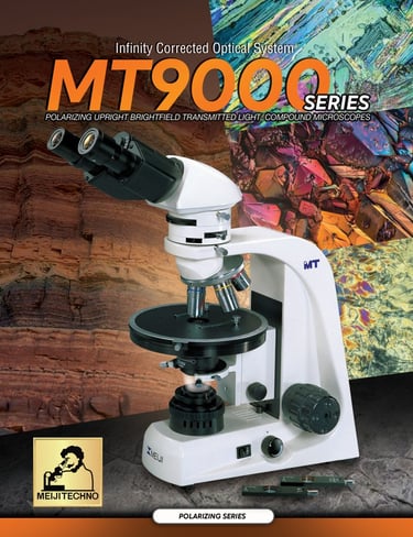
MT9000 Series
The MT9000 Series features include:
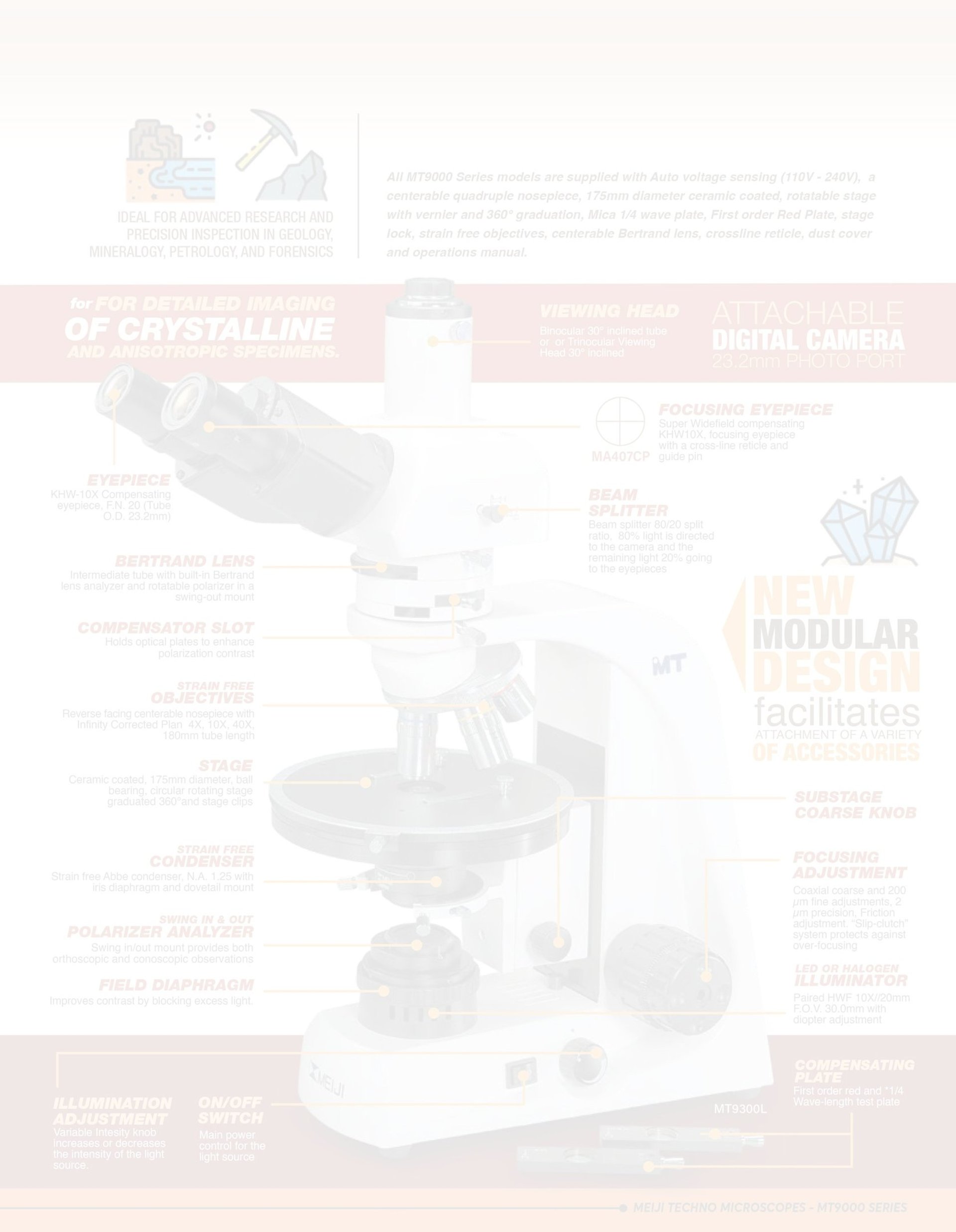
Built in Bertrand lens and analyzer with compensator slots
Swing out Polarizer/Analyzer in sliding mount provides both orthoscopic and conoscopic observations
First order red compensating plate and ¼ Wave-length test in a metal mount sliding mount
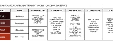
Newly designed Strain free Abbe Condenser 1.25 N.A.
Polarizer in a Swing in-out click/clamp mount, fully rotatable 360° with click stop at 0° and 90°
Super Widefield compensating KHW10X, focusing eyepiece with a cross-line reticle and guide pin
F.N. 20 are standard with 21mm reticle mount
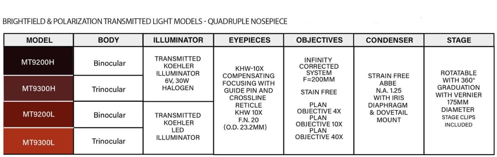
Siedentopf type Binocular or Trinocular viewing heads are standard
Smooth operating ball bearing centerable nosepiece provides effortless objective changes
Clear Observation Images with ? Infinity Corrected Objectives ICOS Optical System
Intermediate tube with built in Bertrand lens and analyzer and compensator slots and rotatable polarizer in a swing-out mount
Ceramic coated, 175mm diameter, ball bearing, circular rotating stage graduated 360°
1° increments and vernier reading to 0.1° circular stage
Focus tension adjustment and safety Auto-focus stage stop lever to prevent slide breakage
Triangular designed frame for maximum ergonomics, greatly lessens user fatigue duringhours of extended observation
Brightfield:
The simplest of all the optical microscopy illumination techniques. Sample illumination is transmitted (i.e., illuminated from below and observed from above) white light and contrast in the sample is caused by absorbance of some of the transmitted light in dense areas of the sample. Brightfield microscopy is the simplest of a range of techniques used for illumination of samples in light microscopes and its simplicity makes it a popular technique. The typical appearance of a brightfield microscopy image is a dark sample on a bright background.
Polarizing Light:
A type of optical microscopy techniques involving polarized light. Simple techniques include illumination of the sample with polarized light. Directly transmitted light can, optionally, be blocked with a polarizer orientated at 90° degrees to the illumination. More complex microscopy techniques which take advantage of polarized light include differential interference contrast microscopy and interference reflection microscopy.
Click on the image to download the brochure

NEC Scientific
Quality laboratory equipment, chemicals, and standards.
info@necscientific.com
+1 (224) 607-1398
© 2025. All rights reserved.